Magnetic Resonance Imaging (MRI) is a revolutionary diagnostic tool that provides detailed images of the inside of the body using strong magnetic fields and radio waves. It is widely used by healthcare professionals to examine almost any part of the body, including the brain, spine, joints, and internal organs. In this article, we’ll take a deeper look at MRI scans, how they work, who can have one, and what to expect during the procedure.
Overview of MRI Scans
MRI scans are one of the critical parts of medical imaging that helps healthcare providers to diagnose, plan treatment, and assess the effectiveness of treatments. They are usually used to look at soft tissues in the body that are not visible using other imaging techniques, such as X-rays.
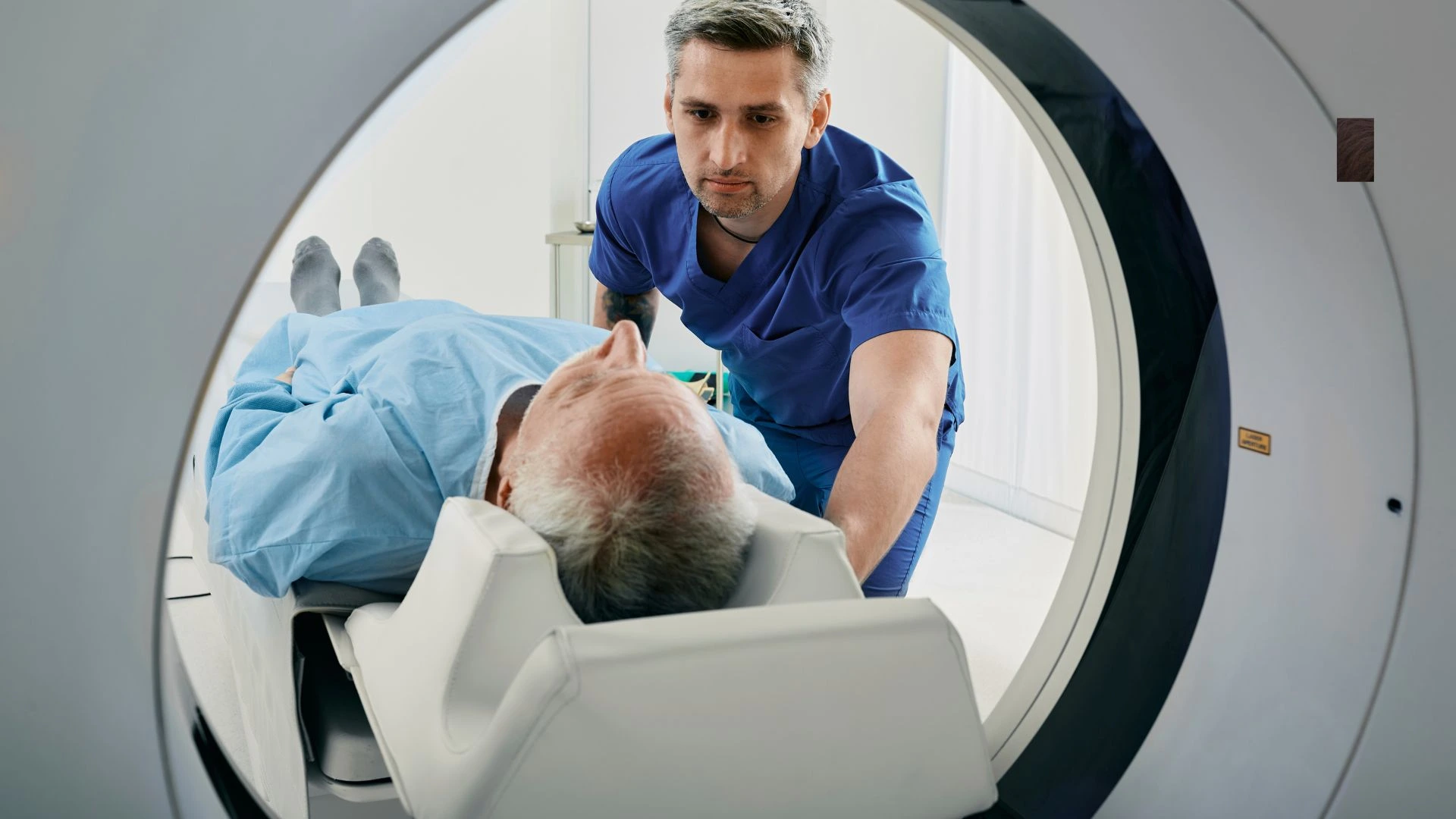
An MRI machine is a large tube that houses powerful magnets. The process involves you lying on a bed that moves into the machine. Inside, strong magnetic fields and radio waves work together to generate highly detailed images of the body’s internal structures. These images can be used to assess everything from brain tumors to joint injuries, helping doctors make informed decisions about treatment.
How MRI Scans Are Performed
The Procedure
This MRI scan is carried out in a specially designed room, normally located in the hospital or in imaging clinics. A radiographer with expertise in medical imaging operates this machine; he controls the machine from another room to save himself from the very strong magnetic field.
You will lie on a flat bed during the scan. The bed moves into the MRI scanner. You will be placed headfirst or feetfirst in the machine depending on the area of your body being scanned. The procedure normally takes between 15 to 90 minutes depending on the size of the area being scanned and how many images are required.
It is essential to be as still as possible during the scan to obtain clear, accurate images. The radiographer may ask you to hold your breath for a few seconds or follow other specific instructions to improve the quality of the scan. You will also be given earplugs or headphones to help protect your ears from the loud tapping noises the scanner makes while it operates.
How Does an MRI Scan Work?
The Science Behind MRI
Water is the primary composition of the human body, composed of hydrogen and oxygen atoms. The proton of a hydrogen atom acts like a small magnet; this sensitivity of protons to magnetic fields explains the basis behind the working principle of an MRI scan.
Upon entry into the MRI machine, a strong magnetic field aligns protons in a given direction within the body. This alignment is then disrupted by the use of short bursts of radio waves. The alignment is reasserted as the radio waves stop and protons return to their original position; in doing so, they give off signals which are then caught by receivers placed within the MRI machine.
The signals produced by those protons are finally used to produce images. Inside the body, there are different types of tissues including muscle tissue, fat tissues, and brain tissues that have associated protons that realign at different speeds. These variations would help in distinguishing between the various tissues and provide clear-detailed images.
Who Can Have an MRI?
Some other conditions and areas of the body that MRI scans are used for include:
Major uses of the MRI scan.
Brain and spinal cord: Mostly, MRI is used to establish neurological disorders involving brain tumors and multiple sclerosis to spinal cord injury.
Bones and Joints: MRI detects damage to bones, ligaments, tendons, or cartilage, like tears in the knee or rotator cuff.
Breast: MRI is used as an additional tool to mammography for women with dense breast tissue to detect breast cancer.
Heart and Blood Vessels: It can examine the heart structure and function, and diagnose diseases in blood vessels, including aneurysms or blockages.
Internal Organs: The use of MRI usually covers the examination of organs like the liver, kidney, prostate gland, and womb.
Doctors may recommend an MRI scan when other imaging tests such as X-rays or CT scans are not detailed enough or in cases where a more precise diagnosis is required.
Who Can’t Have an MRI?
Although MRI scans are relatively safe, there are some situations in which they would not be appropriate. Some conditions or implants could make MRI unsafe. Some of the most common reasons people may not be eligible to undergo an MRI include:
- Metal Implants: People who have metal implants in their body, for instance, pacemakers, artificial joints, or metal clips will not be allowed to have this test since the magnetic field of MRI is extremely strong. It can shift the metal around and interfere with the working of the implant.
- There is no proof that MRI scans pose a threat to unborn babies, yet doctors and other healthcare professionals tend to take precautions. If you are pregnant, your doctor will decide whether you need an MRI and tell you about the risks involved.
- Claustrophobia: In some patients, the feeling of lying inside the closed MRI machine can be associated with anxiety or claustrophobia. The newer machines are designed with wider openings that help in eliminating the feeling of claustrophobia. In case the patient has severe claustrophobia, a sedative may be prescribed or other imaging modalities may be employed.
Safety of MRI Scans
MRI scans are widely regarded as one of the safest medical imaging techniques available. The procedure is non-invasive and does not use ionizing radiation like X-rays or CT scans. Instead, MRI uses powerful magnets and radio waves, which have not been shown to cause harm to the body.
Magnetic Fields and Radio Waves
MRI scanners do produce strong magnetic fields, but they are tightly controlled and localized within the device. The amount of radiofrequency energy used during an MRI is also controlled and kept within limits that are not harmful.
Extensive research has been carried out to gauge the risks posed by magnetic fields and radio waves. So far, no conclusive evidence has emerged to indicate the existence of such health risks if these fields are used for medical imaging. MRI scans are one of the safest procedures in modern medicine.
What to Expect Before, During, and After the MRI Scan
Before the Scan
You may be asked to remove any metal objects, such as jewelry, watches, or hearing aids, before your MRI. These can interfere with the magnetic field and distort images. You might be asked to change into a hospital gown depending on the area being scanned.
If you have any implants, devices, or medical conditions that may impact the procedure, you should tell your doctor and the radiographer. They will help assess whether an MRI scan is appropriate and how to proceed safely.
During the Scan
Lie on the MRI bed, and for this procedure, you have to lie very still. In fact, just as mentioned above, the radiographer is going to work the scanner from another room but will still be able to talk to you over an intercom. It makes a very loud noise, but they will give you earplugs or headphones.
The time required for scanning will vary depending on the area and size of the part to be scanned, from 15 to 90 minutes. You will also be required to hold your breath or stay absolutely still for a certain part of the scan for the images to be sharp and accurate.
Post Scan
You are able to return to your normal routine once the MRI scan is finished. There is no post-procedure aftercare for an MRI scan. You do not have a recovery period. In case you underwent an MRI using contrast dye, your doctor would let you know about any potential side effects.
These images are reviewed by a radiologist who would give a report to your doctor. Your doctor then comes to discuss with you the outcome and further advice in terms of other tests that might be necessary or treatment procedures.
Benefits of MRI Scans
MRI scans provide a wealth of benefits for both doctors and patients:
- Non-invasive and Painless: MRI scans don’t require surgery or injections, making them a non-invasive option for diagnosing various conditions.
- Detailed Imaging: MRI produces incredibly detailed images, making it ideal for examining soft tissues and organs that are hard to see with other imaging techniques.
- No Radiation: Unlike X-rays or CT scans, MRI does not use ionizing radiation, making it a safer option for repeated imaging.
- Early Detection: MRI scans can detect conditions early, even before symptoms appear, which can lead to more effective treatment and better outcomes.
Conclusion
MRI scans are one of the most advanced and safest medical imaging techniques that help to study the inside of the body. The detailed, high-quality images can be crucial for diagnosing and monitoring various health conditions for doctors. Understanding how MRI scans work, what to expect during the procedure, and who can benefit from them is very important for patients who may need this diagnostic tool. Though MRI scans are considered safe, one has to inform the healthcare provider about implants or medical conditions that could alter the procedure. MRI is an important component of modern medicine that performs highly detailed imaging without invasion.


